The presence of anastomosis of two root canals can be easily detected by means of ethylene-diamine-tetraacetic acid gel (EDTA). Its low speed flow consistence together with its opacity permits to visualy detect as it spreads from one root canal to the other. The video shows anastomosis of MB1 and MB2 root canal in the first […]
Autotransplantation
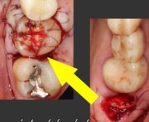
Autotransplantation of left upper third molar to the site of lost right upper first molar. The bridgeworks on both sides were also replaced.
Cervical fillings
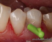
Replacement of failing cervical fillings and demonstration of how to use interdental brush
Recostruction of severly damaged tooth
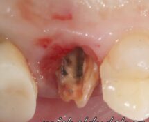
Secondary caries lesion under the metal-ceramic crown and periapical lesion of endodontic origin was addressed during reconstruction of this tooth. In first phase the crown was removed, and composite pre-endodontic build-up was performed. The second phase consisted of root canal preparation, definfection and sealing. In the third phase the tooth core was build-up with fiber […]
Pre-endodontic composite buildup
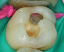
The aim of root canal treatment (RCT) is mechanical and chemical disinfection of the root canal system and its subsequent obturation and sealing. To acquire aseptic operation area it is suitable to isolate the tooth with a rubber dam and properly seal the cavity with a temporary restoration between individual steps of RCT. Since many […]
Minimally invasive treatment of tooth caries
Minimally invasive treatment of tooh caries using tunnel technique. Video of procedure and description of steps (Czech language).
Dental trauma
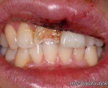
The patient came with broken upper first incisor and contused upper lip. The fracture opened the pulp chamber. The tooth was isolated, the fracture with opened pulp was coated by composite material and the ceramic veneer was fabricated and fixed on the tooth. The dental pulp retained vital.
Dental caries and composite filling
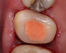
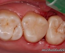
There is carious second upper molar on the first picture. Another caries was found on the front wall of the third molar. The caries on the third molar was treated using priciples of minimal intervention from the frontal approach, to save the most of hard dental tissues and not to weaken the tooth structure. The […]
Plaque
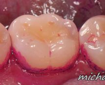
Dental plaque or biofilm is highly organised community of microorganisms, which grow on the surface of teeth. The presence of certain quantity of plaque on the teeth is perfectly normal and the substances it produces are neutralized by saliva, but in the case of excessive plaque accumulation it causes damage to teeth, gums and bone. […]

